


Onychomycosis is a type of fungal lesion only affecting the surface of the nail.To prevent the spread of infection in time, you need to know what nail fungi is, because the therapeutic effect starting in the early phase of the infection will be achieved faster.Most diseases occur in older people due to poor immunity, but the risk of taking illness refers to every person.There is no classification of cork nail damage;In medical practice, it is customary to distinguish the depth of localization and the depth they have penetrated.The infection is also grouped depending on the type of pathogen.
Types of pathogens and primary signs
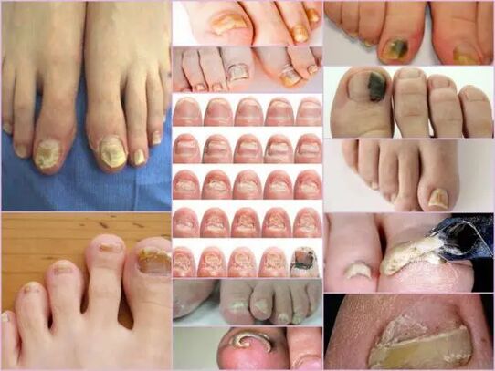
The symptoms of nail fungi in the initial stage are easily identified with the condition of the nail.This sign is the most informative, because onychomycosis always shows itself in the form of change in the color of the surface, in deformation, wear and any external changes.The last, the formation of rudeness, grooves and cracks, as well as the point of intersection, and the point is expressed by the general violation of the nail.
The main sign of a healthy nail is pink and transparency.At any stage, it is characterized by the bulging of the monkomycosic nail and to yellow, brown or green with color change.In an advanced stage, the surface can get a black shade in the background of almost completely destroyed.
The external signs of infection with a mushroom depend on the type of pathogen.The following possible lesions differ in medical practice:
- With a mushroom of Candida, with a mushroom directly from the box with the discharge of the nail.Candidates of rolls around the nail will be characteristic of the candidiasis.This version of the complication may have a bacterial source in the form of either streptocococci or staphylococci or be expressed in the form of a medium or psoriasis plate;
- Dermatophites of Trichophyton Rubrum type.In this case, the reputation of the infection occurs directly from the free edge of the nail.The first symptoms of such a pathogen are the appearance of a yellowish point on the surface.Neoplasm nails crashes and the ground itself tends to increase.The general location of neoplasm's localization, parallel to nail reins, along the plate, is called distal-lateral infection.This pathogen has another form of defeat - distally distal, distally visible in the earth, mainly in the middle of the free edge;
Important!
These types of fungi are in the thumbs of the toes of the legs, make the nails a blank mass.In the absence of proper treatment, the disease runs hyperkeratose.The nail plate is completely destroyed due to the spread of infection around the perimeter.
- Dermatophytes like Trichophyton Mental Cauldrophytes.With onychomycosycosy, such excitements are also smaller than small fingers, most thumbs nails.This type requires an infection with a fungus, not just nail, but also the legs due to rapid spread of pathogen.A symptomatic disease can be like Leikonichia, a common disease in medical practice.Primary signs are white spots visible in different parts of the nail, neoplasms differ in unstable shape and different sizes.Leakonichia's fungi is easy to distinguish - in the last case, the spots are the accumulation of air not observed in mushroom damage;
- Mushroom mushrooms.This damage option is significantly less than a namandal or dermatophyte form.It just takes the main sign of such an infection - the surface of the plate, almost black shade.All your finger cannot be completely infected, but part of the nail plate.The first signs of nail mushrooms on this type of legs are a sharp change in color.Onychomycosis can develop in the form of a black or dark green in color in color in color in the background of the rest of the pink section of the nail.
Diagnostics by type of change
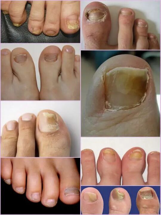
It is not difficult to feel a nail fungus on the main stages of this type of infection, because this type of infection is actively actively actively active in the first day of the lesion.Instead of ordinary transparent nails, which are pale pink in the patient, a significant surface deformation and conditions are observed in the conditions.The affected area has a matte boring yellowish color that is mainly visible in the thumbs.The type of mushrooms and damage to the damage are determined.
In the first stage, the feet of the legs in the legs are the appearance of a small focus.The thickening of the slab and the ceratinating of the bed under the nail will be characteristic.This stage is accompanied by a partial separation and discharge of the nail plate that serves as a source of infection for healthy people by such a phenomenon.
Despite the active thickening of the tile, permanent grinding can be observed, regardless of the present factors.The features of each stage and the symptoms of fungi depend on the legs directly to the pathogenic type.
Depending on the changes occurring with nail plate, three options for onyokomycosis are different:
- Hypertrophic;
- Normotrophic;
- Atrophic.
In the first case, a sharp change in the shadow of the nail plate, destroying along the edges, as well as deform the surface of the plate.The nail is so thick that causes anxiety and painful feelings during a walk.Normotropic micosu on the legs in the legs distinguish with a healthy glow, but the plate itself is a slight hug and placed on it.With an atrophic damage, brown and gray focal form on the surface of the nails.Thanks to them, it is possible to clearly define the location of the pathogen.
Important!
Atrophic or checked body type nail fungi, as in other cases, are characterized by the finishing of the nail plate, as in other cases.The areas where the pathogen are localized tend to leave.The absence of proper treatment leads to an advanced stage - a complete rejection of the nail plate.
Localization classification
Another sign that your toes of the toenails can separate the damage to the nails depends directly on the spot of the focus on the nail plate.This also includes a depth of pathogen, which in turn allows you to determine the approximate period of the upcoming therapy and a speedy chance of recovery.
Mushrooms of the legs of the legs in localization are classified into the following groups:
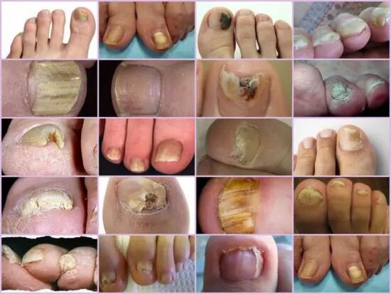
- Onychomycosis is a white surface type - the appearance of many white spots on the surface of the nail plate.A fungal infection causes skin to separate the skin in the appearance of the skin of the skin.The advanced stage causes the full destruction and rejection of the plate;
- Distal - develops the free edge of the nail.The color change is initially observed in a small area of the plate, and then it has an active expansion of its borders.The lesion is characterized by a ton of yellowish or brownish color, but also a rough uneven surface and gradual abrasion;
- There are stages of distalance in lateral Onychomycosis, but only localized on the sides of the nail plate;
- Common infection - full infection of the entire surface;
- Proximal onychomycosis.This disease begins with the activities of the Cuticle, which is inflamed, then the mushrooms quickly affect the nail, and the process itself begins with a small white spot near the inflammatory territory of the perivious roll.
The most common forms of micosing nails in the feet are lateral and distal to cause dermatophyte.The forms of damage such as proximal and white can act as a secondary disease that accompanies the disease of the disease of the vich.The cost of the round with a mushroom should be regarded as a stage of development of each fungus under the nail.
Features of fungi under a microscope
Despite the effective number of Onychomycosis, other diseases related to skin problems and other non-contagious diseases in nature are taken to damage the legs of the feet.Only under one microscope, you can identify the exact diagnosis and type of pathogenic, for which the biological material is being crumbed in the hospital.
The biomerial, in a alkali environment, was already changed in advance, and after which a lot of growth is seen.The study of nail mushrooms under a microscope allows you to see an external shape, external shape, which determines its type, distribution scale and approximate damage.In a nutritious environment, you can only predict the approximate increase of colonia, which allows you to make a clear diagnosis, but also to determine the restriction of infection.
Important!
Since Onychomycois is possible to determine the presence of only in a laboratory, you should not postpone a visit to the doctor with the slightest doubt.Nail fungus is growing rapidly and the loss of time increases therapy.
What is a malicious signal?
The mushrooms of the nails on their feet show themselves with a number of similar symptoms in some skin diseases.The following symptoms are more likely to indicate the Onychomycotic lesion that requires medical intervention:
- The appearance of the yellow spots in the plate, its deformity, a change in the previously observed structure;
- Demonstrates a shadow of black color, which is characteristic of the nail mushrooms on the blackspace of the tiles, loss of transparency, the black color characteristic for the molds of pathogens;
- The thickization of the nail plate and a very sharp thinning of the keratinization or vice versa;
- Bunch of bits, scales and crumbs from the nail;
- The swelling of the wheel hangs over the plate, leaves liquid;
- Regardless of the type of pathogen, the people of the fungi are visible nails.
All these symptoms are high at risk of infection.Some external symptoms change the structure of the nail plate can be the result of other diseases.With the increase in fragility, the amount of calcium and iron in the body must be increased, which means any infection of the growing body to grow, purely white stripes and dots, copper or zinc deficiency.Although the photograph, nail fungi, which is a change in the plate, which is a change near the yellow color, it does not always show infection.The jaundice can show various problems with the stomach and liver.
Despite the extensive classification of the disease, it is not difficult to recognize onychomycosis on the fingers with external signs.However, you can determine the exact type of pathogen and the damage phase can only be in the laboratory.